Welcome to the Department of Functional Diagnostics at Kimyo University Hospital
At Kimyo University Hospital - we provide state-of-the-art medical services across all fields of medicine, and our Functional Diagnostics Department is no exception. Our department offers a comprehensive range of diagnostic imaging services to visualize internal body structures. This includes imaging of the chest, bones, joints, and soft tissues, enabling the detection and diagnosis of fractures, infections, tumors, and other diseases. At Kimyo University Hospital, we prioritize patient comfort, safety, and confidentiality.
Our diagnostic services include advanced techniques such as CT angiography, which allows for detailed visualization of blood vessels in areas like the thoracic aorta, abdominal aorta, and upper and lower extremities. We also perform three-phase studies of the kidneys and abdominal cavity to provide thorough and precise diagnostic information.
By leveraging cutting-edge technology and a patient-centered approach, we ensure the highest standards of care in medical imaging and diagnostics.
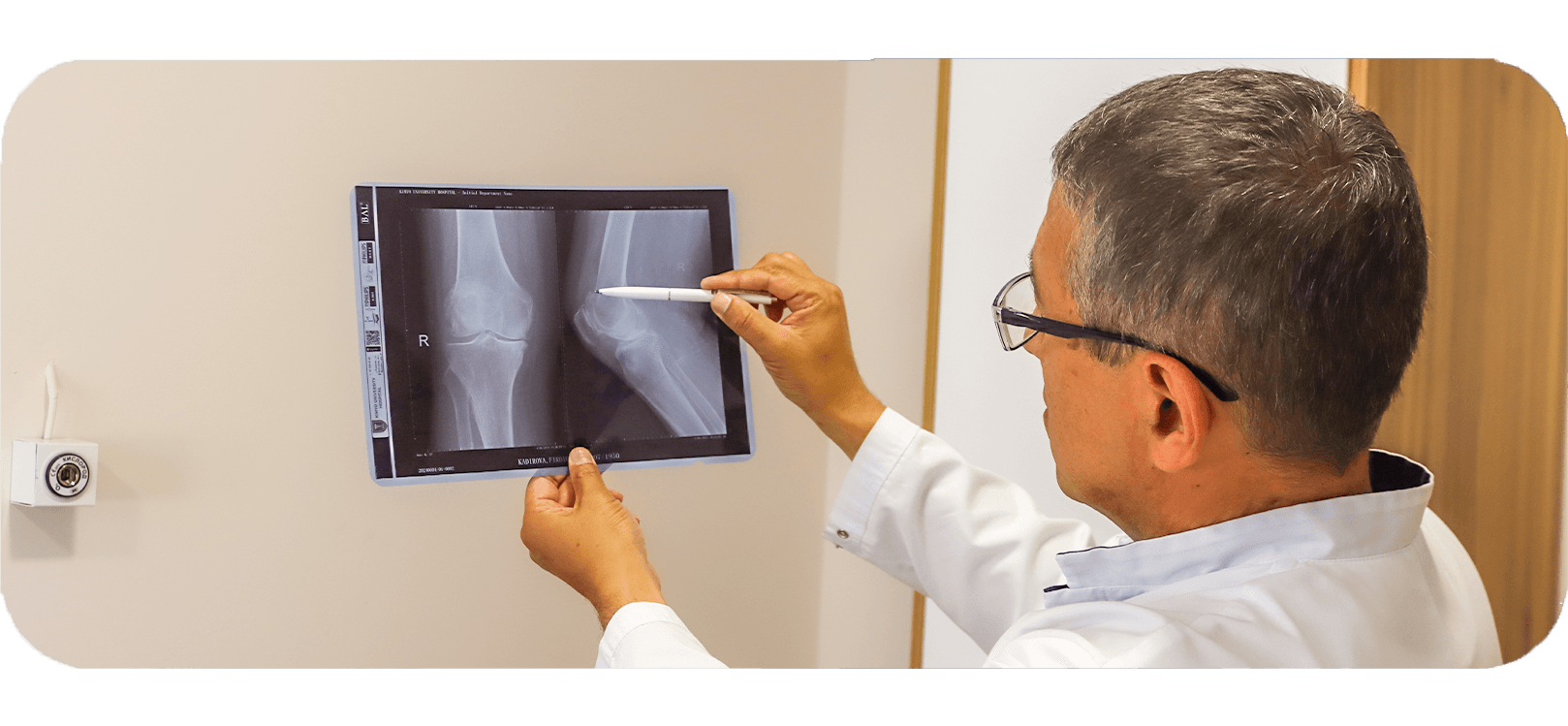
Radiography - is the study of the internal structure of objects that are projected using x-rays onto a special film or paper.
At Kimyo University Hospital, this includes imaging of the chest, bones, joints and soft tissues to detect and diagnose fractures, infections, tumors and other diseases. At Kimyo University Hospital, we prioritize patient comfort, safety and privacy.
Our services:
Chest X-ray
Dental X-rays
Abdominal X-rays
Bone X-rays
Mammography
Spine X-rays
Fluoroscopy
Welcome to Kimyo University Hospital's Department of Ultrasound!
At Kimyo University Hospital, we offer cutting-edge medical services in all spheres of medicine, and our Department of Ultrasound is no exception. Ultrasound, also known as sonography, is a non-invasive imaging technique that uses sound waves to create real-time images of the internal structures of the body. This technology allows us to visualize organs, tissues, and blood vessels, aiding in the diagnosis and monitoring of various medical conditions. At Kimyo University Hospital, we prioritize patient comfort, safety, and confidentiality.

Angiography is a method of X-ray contrast examination of blood vessels, often used to diagnose various diseases. This method allows visualization of vessels in any part of the body, including the head, limbs, and heart. Angiography helps evaluate the presence and location of both normal and accessory arteries; determine the degree of preservation of their lumen, identify narrowings (stenosis) or expansions (aneurysms) that can disrupt blood flow; and also examine the structure and integrity of vessel walls and the quality of blood flow. This method is widely used in the diagnosis of cardiovascular diseases, strokes, aneurysms and other vascular pathologies.
CT scan angiography of the thoracic aorta
CT scan angiography of the abdominal aorta
CT scan angiography of the Upper and Lower extremities
Three-phase study of the kidneys and abdominal cavity.
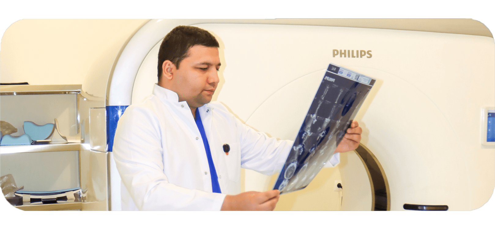
CT scan (Computed tomography) is a study in which a tomograph takes many X-ray images that are processed by a computer to obtain a complete picture of the internal organs. The procedure is safe for adults but is contraindicated during pregnancy due to the high level of radiation exposure, especially in the first trimester when the baby's major organs are forming. Even when examining the extremities or head of the expectant mother, minimal radiation exposure can disrupt the development of the fetus, which may manifest itself in adulthood.
В отделении эндоскопии Kimyo University Hospital проводятся современные малоинвазивные методы диагностики и лечения заболеваний желудочно-кишечного тракта, дыхательной системы и желчевыводящих путей. Все процедуры выполняются высококвалифицированными специалистами на оборудовании последнего поколения.
ЭГДС позволяет визуально оценить состояние слизистой оболочки пищевода, желудка и двенадцатиперстной кишки. Применяется при жалобах на изжогу, боли в желудке, тошноту, рвоту, анемию и при подозрении на язвы, гастрит или опухоли.
Специализированный осмотр двенадцатиперстной кишки, часто используется в комплексе с ЭРХПГ для оценки проходимости желчных протоков и выявления опухолей, стриктур или камней.
Позволяет осмотреть всю толстую кишку с целью диагностики полипов, воспалений, опухолей, язвенного колита, болезни Крона и других патологий. Часто назначается при длительных болях в животе, нарушениях стула и скрытых кровотечениях.
Менее обширное исследование, направленное на осмотр сигмовидной и прямой кишки. Часто используется для экспресс-диагностики при жалобах на кровь в стуле, боли внизу живота или подозрении на опухоли прямой кишки.
В случае выявления подозрительных участков врач может взять образцы ткани для гистологического анализа (биопсия), чтобы исключить онкологические и предраковые состояния.
Диагностическая процедура для осмотра трахеи и бронхов. Применяется при длительном кашле, подозрении на опухоли, бронхиальных кровотечениях, хронических инфекциях или при необходимости удаления инородных тел. Возможна бронхоскопия с забором материала (биопсия).
Удаление доброкачественных новообразований — полипов в желудке или кишечнике. Это помогает предотвратить развитие рака, особенно в случае аденоматозных полипов.
Удаление ограниченных поверхностных поражений слизистой желудка или кишечника. Подходит для начальных стадий рака без глубокой инвазии.
Более точная и глубокая методика удаления ранних опухолей. Позволяет полностью удалить новообразование без открытой операции.
Безопасное извлечение случайно проглоченных предметов у детей и взрослых с помощью гибкого эндоскопа.
Создание доступа для питания у пациентов, неспособных самостоятельно глотать (например, после инсульта или при раке горла).
Применяется при сужениях пищевода или кишечника (стриктурах), вызванных воспалениями, операциями или опухолями. Позволяет восстановить проходимость пищеварительного тракта.
Наложение резинок на варикозно-расширенные вены для предотвращения и остановки кровотечений при портальной гипертензии.
Введение специального вещества в варикозные вены пищевода, вызывающее их закрытие. Метод используется при активных кровотечениях.
Врач может применить клипсы, электрокоагуляцию или инъекции для эффективной остановки желудочно-кишечных кровотечений.
Малоинвазивная процедура: в желудок через рот устанавливается силиконовый баллон, который заполняется физиологическим раствором. Он уменьшает объем желудка и вызывает быстрое насыщение. Подходит пациентам с ожирением I, II и III степени, особенно при отсутствии эффекта от диет, физических нагрузок и других консервативных методов.
Инъекция ботулотоксина в стенку желудка замедляет опорожнение и снижает аппетит, что помогает сбросить вес. Метод рекомендован пациентам с ожирением I–II степени и может использоваться как дополнительная мера в комплексе с программой питания и физической активности.
Создание сужения желудка с помощью эндоскопических швов. Позволяет уменьшить объем съедаемой пищи без хирургического вмешательства. Эффективна и безопасна при ожирении I и II степени, а также может применяться как подготовительный этап перед бариатрической операцией при ожирении III степени.
Комбинированная процедура, позволяющая диагностировать и лечить заболевания желчных и панкреатических протоков.
Контрастное вещество вводится в протоки для получения рентгеновского изображения и уточнения диагноза.
Через эндоскоп камни извлекаются из общего желчного протока без необходимости операции.
Малое рассечение мышечного кольца (сфинктера Одди) для облегчения выхода желчи и доступа к камням.
Металлические или пластиковые стенты вводятся в протоки при опухолях или сужениях, чтобы восстановить отток желчи.
Извлечение объектов, случайно попавших в дыхательные пути (у детей или пожилых людей).
Помогает при тяжелых формах бронхита, пневмонии и обструкции дыхательных путей.
Промывание бронхов физиологическим раствором с диагностической или лечебной целью (например, при инфекции или аспирации).
При сужении дыхательных путей (опухоли, рубцы, сдавление) вставляется стент для восстановления проходимости.
Изоляция определенного участка лёгкого (например, при фистулах или локальных кровотечениях).
Временная остановка массивного кровотечения с помощью специальных приспособлений или введения тампонов.
Мы предлагаем полный спектр диагностических и лечебных эндоскопических процедур в соответствии с международными стандартами. Команда квалифицированных врачей и современное оборудование позволяют точно диагностировать и эффективно лечить широкий круг заболеваний.
MRI is a modern and highly informative diagnostic method that allows detailed visualization of internal organs and tissues without the use of ionizing radiation. The examination is performed using a magnetic field and radio waves, making it safe and suitable for repeated use when necessary.
At our center, examinations are performed using the uMR 670 (1.5T) scanner — a next-generation MRI system that provides image quality comparable to 3.0T systems while ensuring a high level of patient comfort.
The system is equipped with advanced image processing technologies, including AI-based reconstruction (DeepRecon), which improves image clarity and detail. This is especially important for detecting subtle pathological changes at early stages.
Optimized scanning protocols reduce examination time, minimizing motion artifacts and improving patient comfort.
The wide 70 cm bore and ergonomic design help reduce anxiety and claustrophobia, which is particularly important for children, elderly patients, and those with pain.
EasySense technology enables respiratory monitoring without external sensors, simplifying the procedure.
QScan technology reduces acoustic noise, making the scan more comfortable.
High system performance ensures stable image quality and allows complex diagnostic protocols.
MRI of the head
Brain MRI
Cerebral vessels MRI (MR angiography, MR venography)
Brain MRI with сосуды
Pituitary MRI
Brain and pituitary MRI
Brain and orbit MRI
Paranasal sinuses MRI
Brain MRI with epilepsy protocol
Brain and inner ear MRI
Cranial nerve MRI
Brain MRI with tractography
MRI of the spine
Cervical spine MRI
Thoracic spine MRI
Lumbosacral spine MRI
Sacrum and coccyx MRI
Spine MRI with myelography
Whole spine MRI
MRI of joints
Temporomandibular joint MRI
Shoulder joint MRI
Elbow joint MRI
Wrist joint MRI
Hand MRI
Hip joints MRI
Sacroiliac joints MRI
Knee joint MRI
Ankle joint MRI
Foot MRI
MRI of internal organs
Chest and mediastinum MRI
Abdominal MRI
Abdominal MRI with cholangiography
Pelvic MRI
MRI of soft tissues
Neck soft tissue MRI
Soft tissue MRI of various regions
Extremity soft tissue MRI
MRI with contrast
Brain MRI with contrast
Pituitary MRI with dynamic contrast
Internal organs MRI with contrast
Spine MRI with contrast
Soft tissue MRI with contrast
MRI angiography
Cerebral vessels MRI
Neck vessels MRI
Major and extracranial vessels MRI
Comprehensive studies
Whole spine MRI
Half body MRI
Whole body MRI